Brasil
 Global
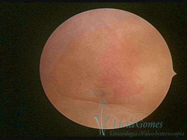
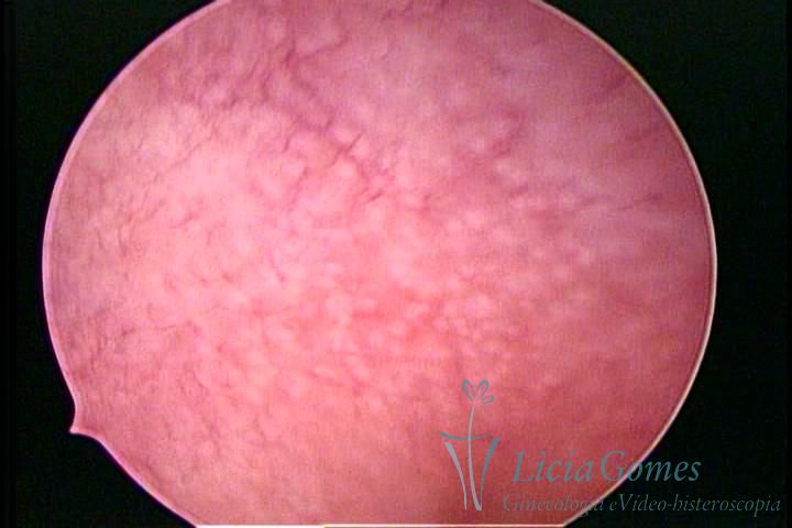
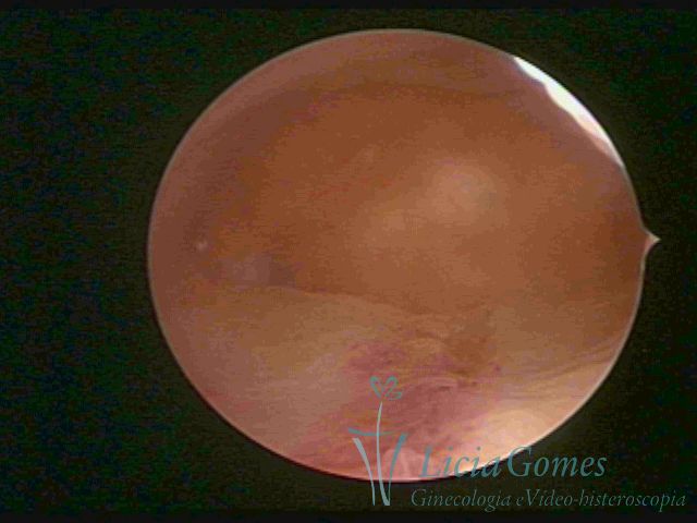
NORMAL UTERINE CAVITY
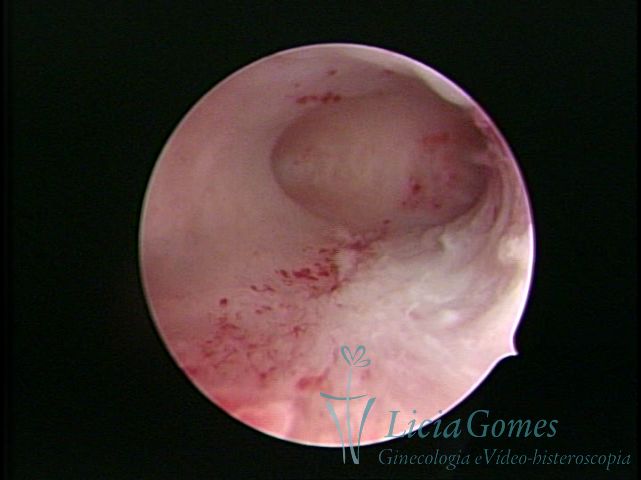
From the internal orifice, the distended uterine cavity is presented in the shape of an inverted cone, whose base smoothly dilates to a point where it opens more, presenting, from this point, an ovoid shape with a larger transverse diameter. On the extreme borders, the cornual regions are excavated and mouthed in a divergent slanting course to the tubal ostiums.
The conical shape constitutes the segment or lower part, and the ovoid structure, the segment or upper part of the uterine cavity.
The normal uterine walls are homogeneously distended, precisely defining the two segments and the cornual excavations.
The normal endometrium covers the whole surface, from the internal orifice of cervical canal to the tubal ostiums.
Menstrual cycle may be defined as the time between the beginning of menstruation to the beginning of the subsequent.
In this time, the endometrium, subject to the actions of the hormonal cycle, changes and, in order to present a better comprehension, the hormonal cycle has been divided into phases (it is important to standardize the changes of the endometrium during the menstrual cycle in order to compare the possible inflammatory or hormonal changes or drug action.
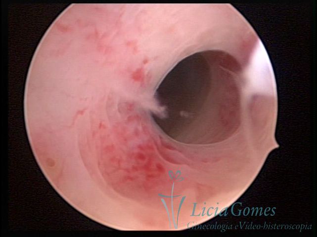
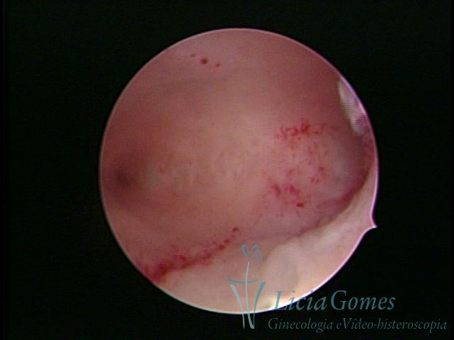
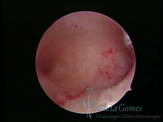
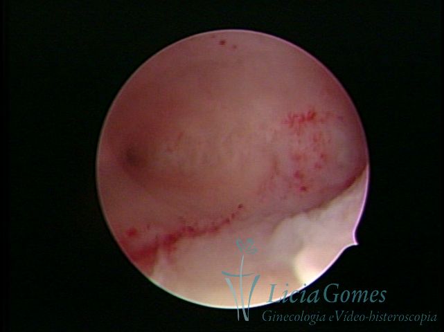
Day 1: Initial menstrual endometrium: acquires an intense red color, presents deep hemorrhagic crackings, extense superficial hemorrhages and congestion. The surface is irregular, alternating practically exposed zones with other zones where the mucosa is preserved.
Days 2 to 4: Final of menstruation: the endometrial surface presents a red coloration - which is intense due to small hemorrhagic spots. The uniform surface and minimal roughness are noted. Occasionally, is possible to observe white-colored endometrial accumulations in hemorrhagic zones and/or authentic endometrial stripes between the growing mucosa zones, which present a purplish color.
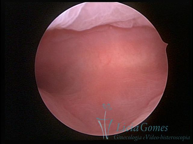
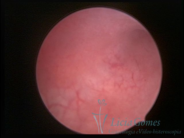
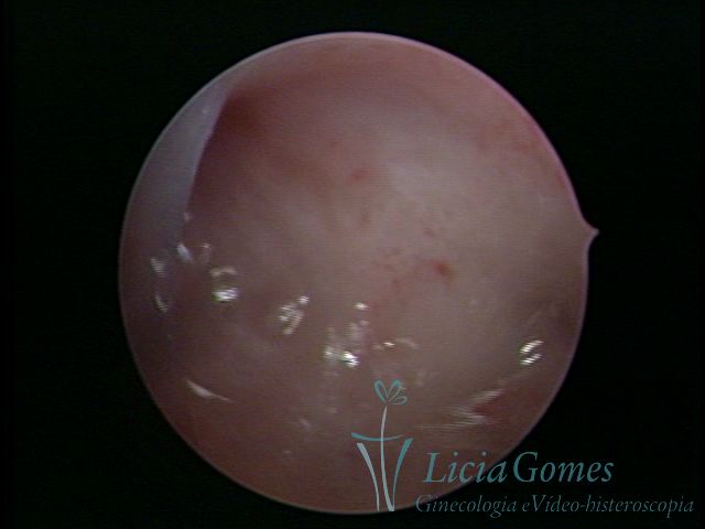
Days 5 to 10: Initial proliferative phase: the endometrium presents an amber-like color. The surface is flat, smooth and uniform, with an approximate thickness of 2-5mm. With the view of the white spots (endometrial glands.) The vessels present the shape of small, red spots with the shape of small, thin, frequently interrupted capillaries. As the proliferative phase progresses, the vessels become more visible with a more accentuated passageway and acquire an irregular distribution. The cornual regions are well excavated and have a shape of a funnel, since there is not the formation of functional ostiums from the endometrium yet.
In the more advanced proliferative phase (Days 10 to 14 of the Cycle,) the endometrium becomes thicker, with a smooth, shiny surface of a red or yellow coloration. The glands appear as sparse white spots. The largest growth of the endometrium decreases the depth and the tapering grade of the cornual excavation.
Days 11 to 15: Advanced or late proliferative phase: endometrium. It presents a smooth, shiny surface of a red or yellow coloration. The glands appear as sparse white spots with a 6-7 mm thick. The endometrial glands can be clearly observed. The superficial vascular system presents thin vessels with a well-defined passageway.
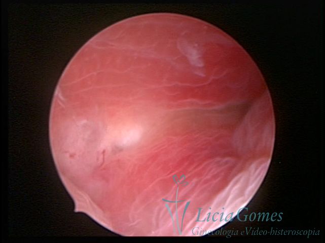
Days 16 to 18: Ovulation phase (initial secretory phase:) the uterine walls are more distended due to the relaxing action of progesterone, which provides a more enlarged aspect to the cavity structure. The endometrium is quite thick, presenting a red coloration and superficial drops which may vary from light swells to bossings separated by accommodation folds. The gland orifices are presented as pales spots with a thin surface and sparse and irregular distribution. The superficial vascularization outlines the capillary system which demarcates the tubal orifices. The tubal ostiums are thinner, more separated from the muscular cornual apex which provides a more shallow concavity. In the advanced secretory phase (days 20 to 25 of the cycle) the endometrium may reach 6-8mm. It acquires a stronger red coloration. The surface may present drops which resemble dunes, bossings, or polypoid and short digitations by stromal edema. The cornual regions are more shallow and the functional tubal ostiums are punctiform, delimited by a thin, shiny circular skin called pre-tubal diaphragm.
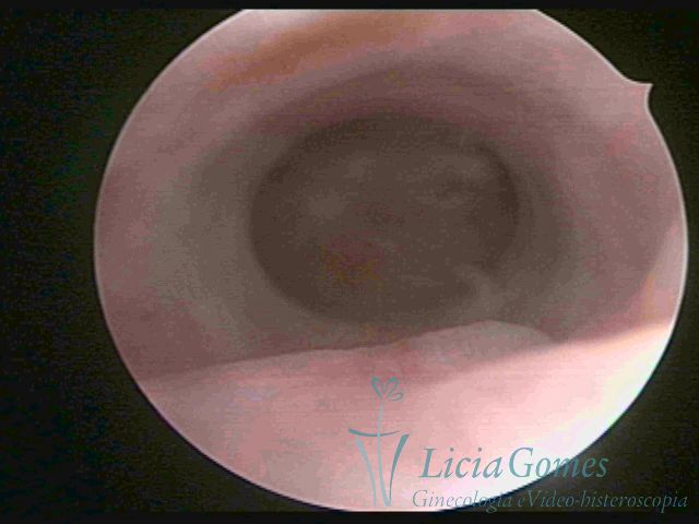
Days 26 to 28: First signs of menstruation: the endometrium presents an opaque, white gray coloration due to the disappearance of the edema. The surface is flat and the endometrium is more compact. Zones with thin excavations and hemorrhagic spots on the endometrial surface may be observed. The thickness is lower than 7mm. Glands or vessels are not observed.
Immediate premenstrual endometrium: the surface presents several hemorrhagic, deep crackings, which confers the cerebroid aspect followed by remote, gross hemorrhagic spots. In zones with a greater retraction, the presence of blood may be observed.
 1
1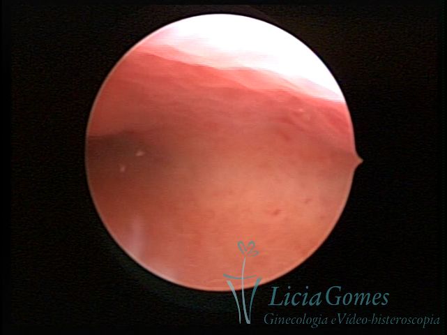 2
2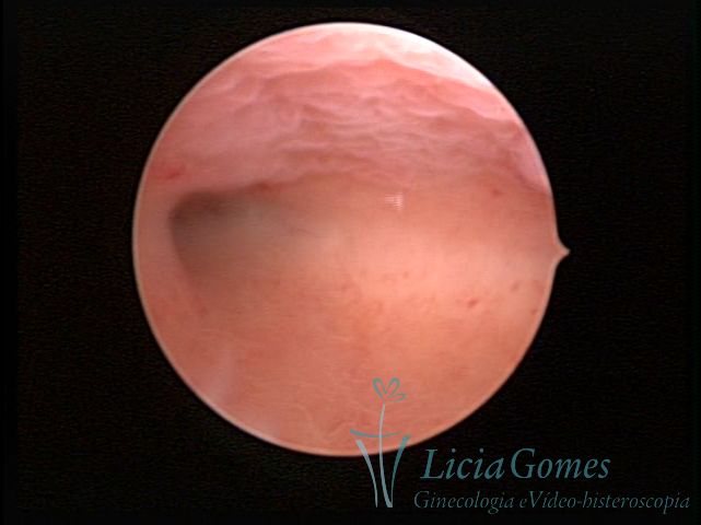 3
3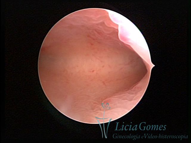 4
4 5
5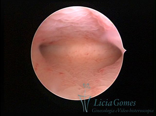 6
6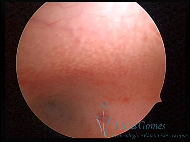 7
7 8
8 9
9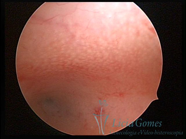 10
10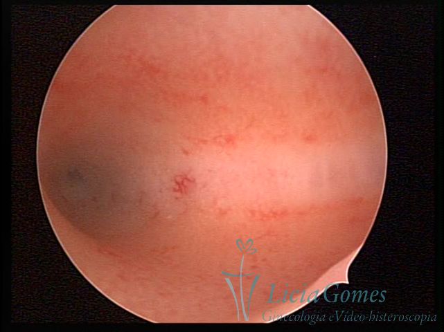 11
11 12
12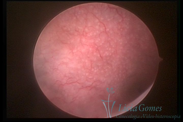 13
13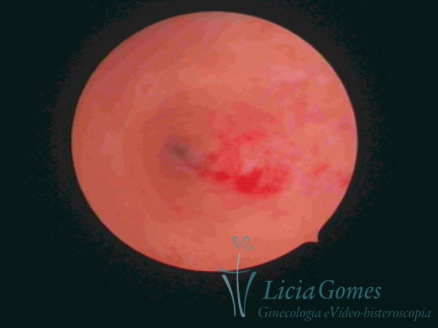 14
14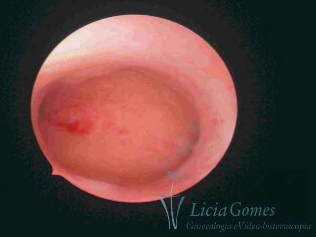 15
15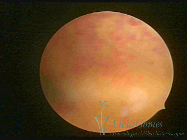 16
16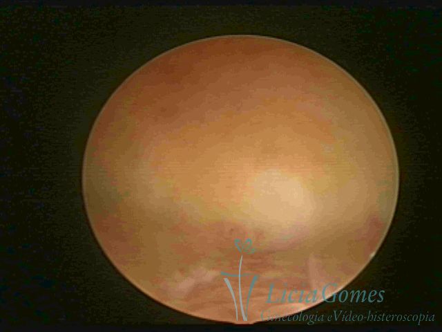 17
17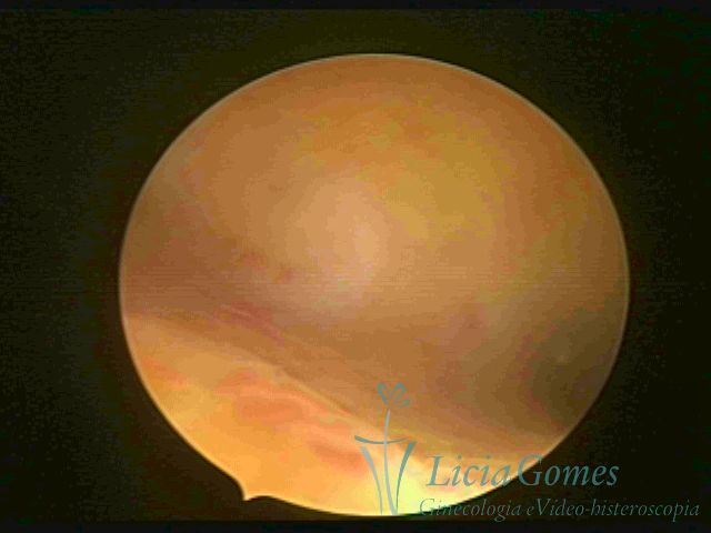 18
18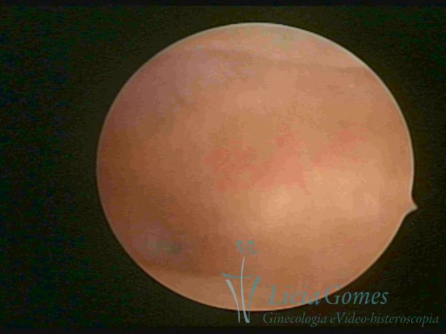 19
19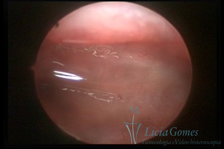 20
20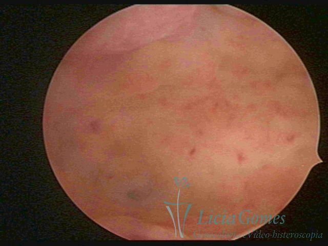 21
21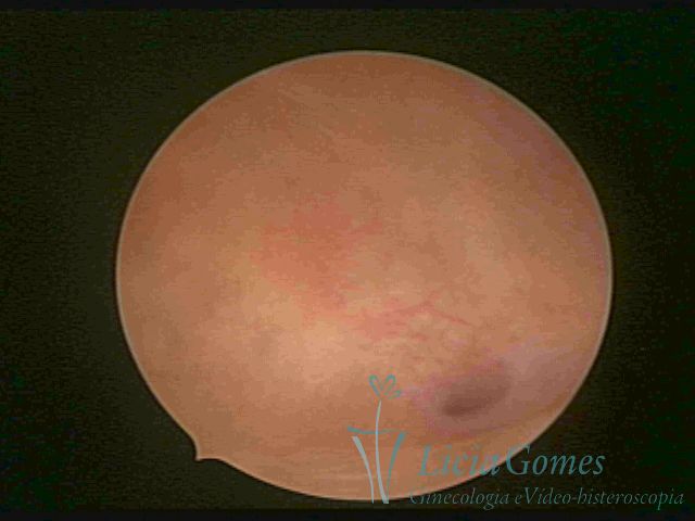 22
22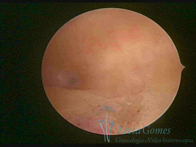 23
23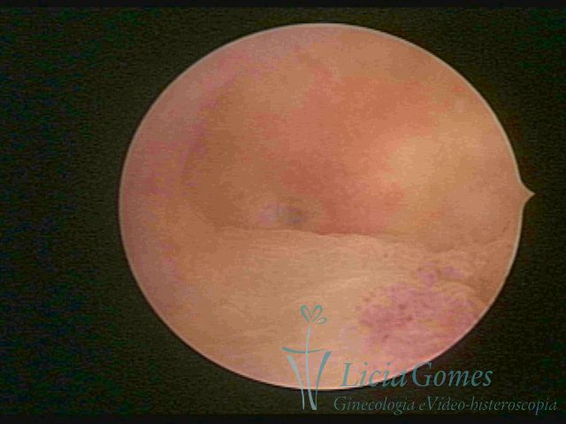 24
24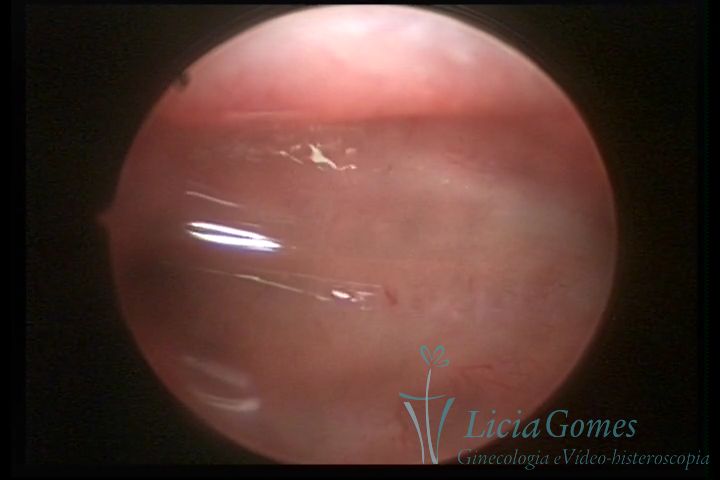 25
25 26
26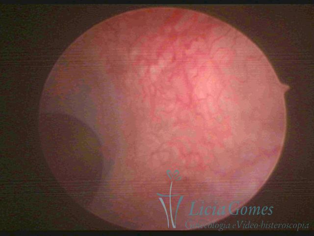 27
27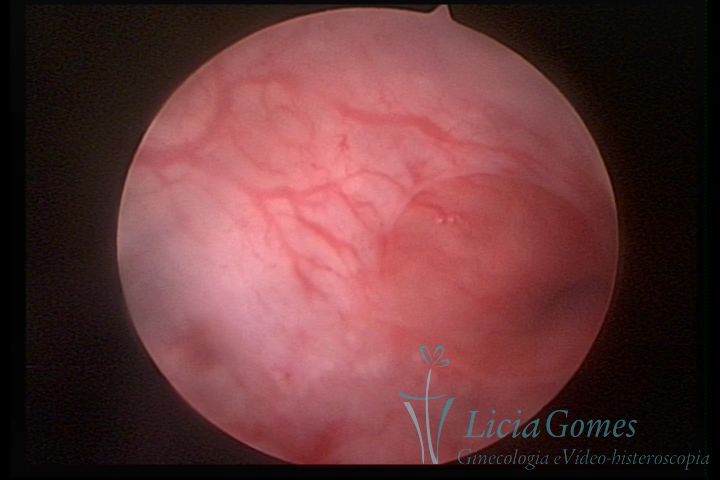 28
28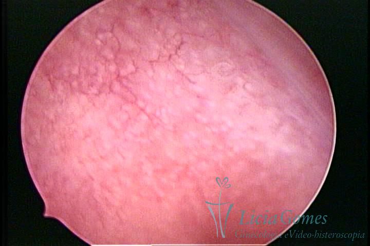 29
29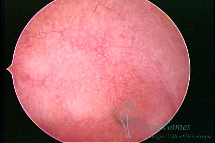 30
30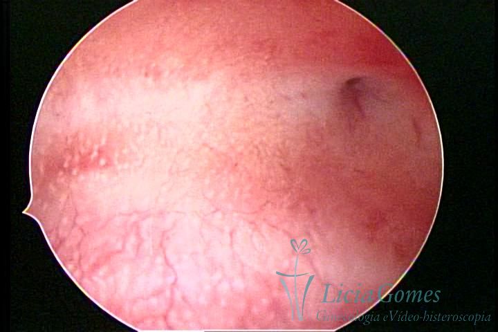 31
31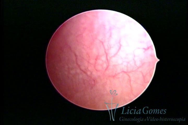 32
32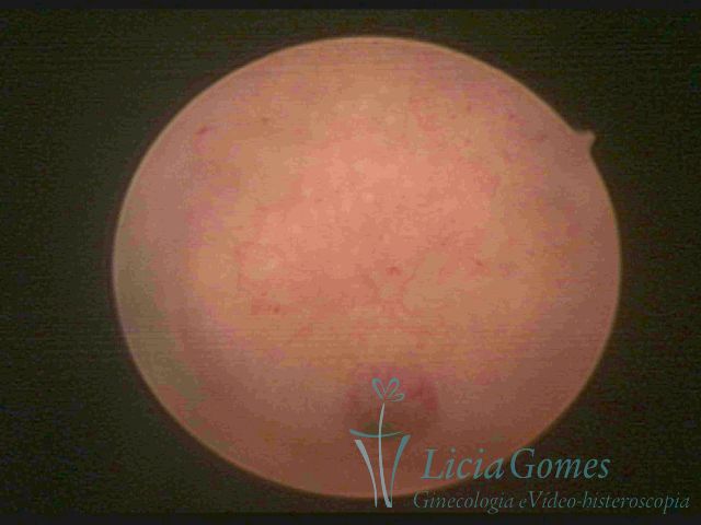 33
33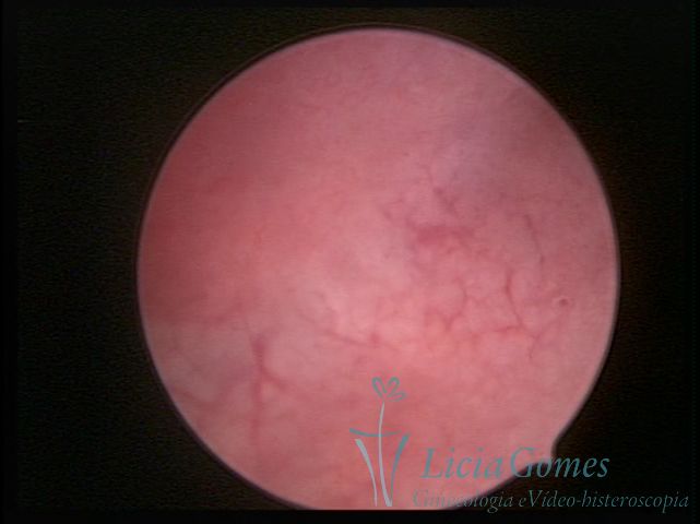 34
34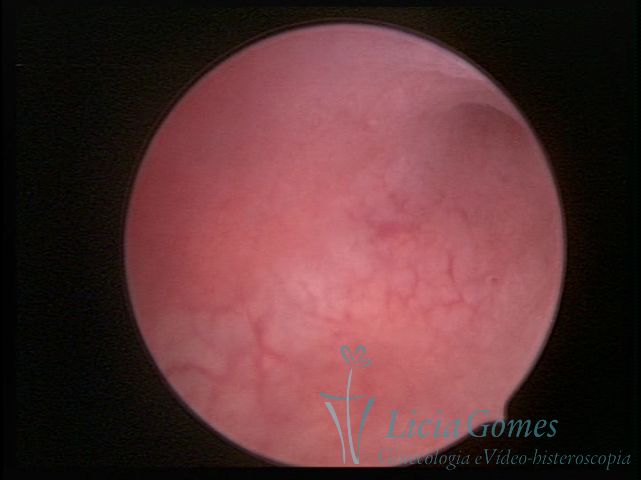 35
35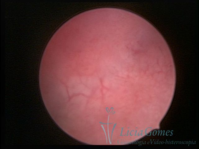 36
36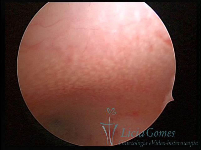 37
37 38
38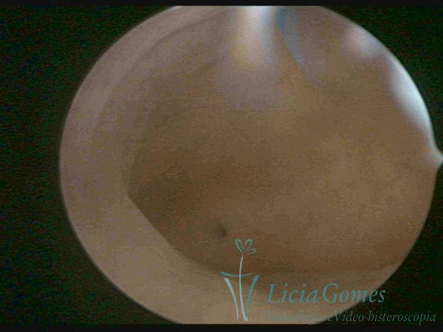 39
39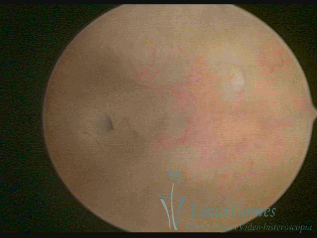 40
40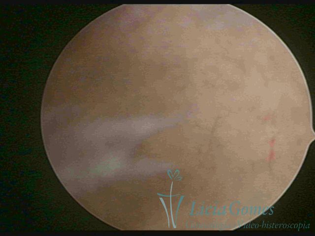 41
41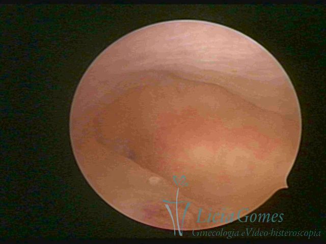 42
42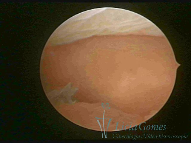 43
43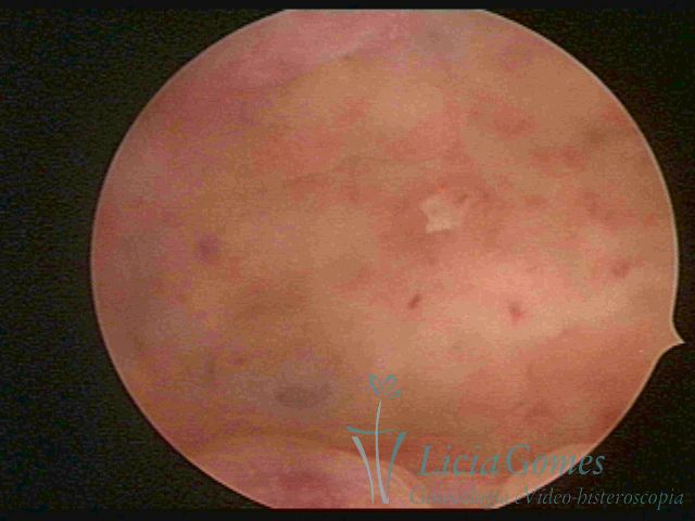 44
44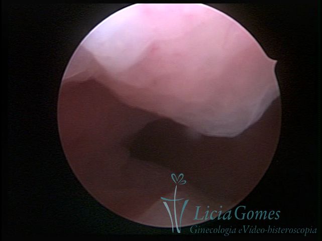 45
45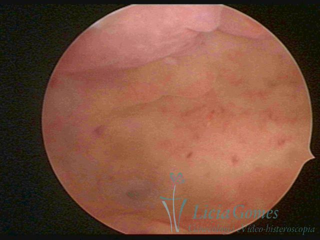 46
46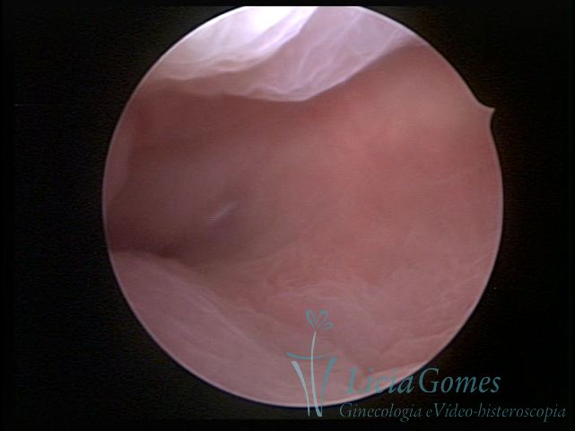 47
47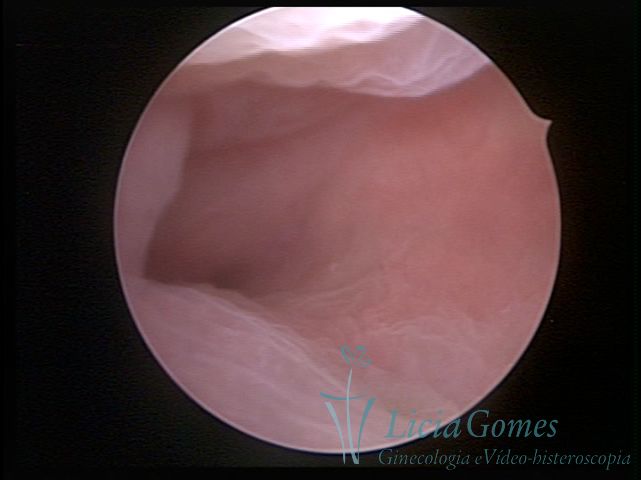 48
48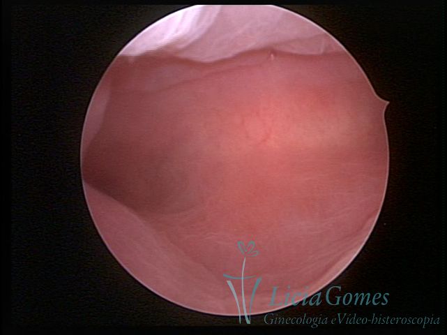 49
49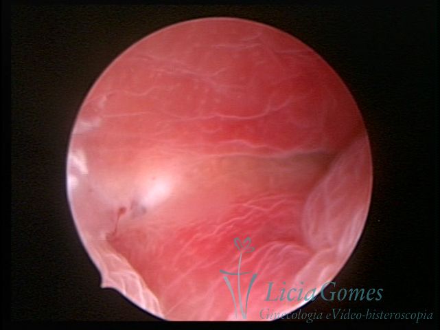 50
50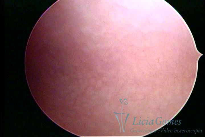 51
51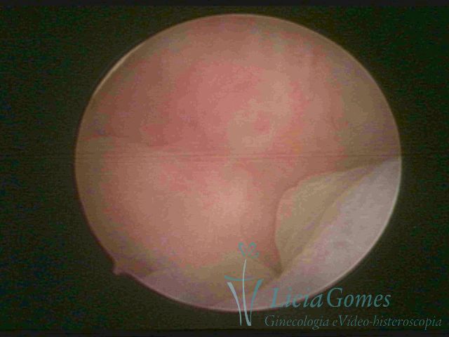 52
52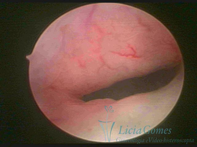 53
53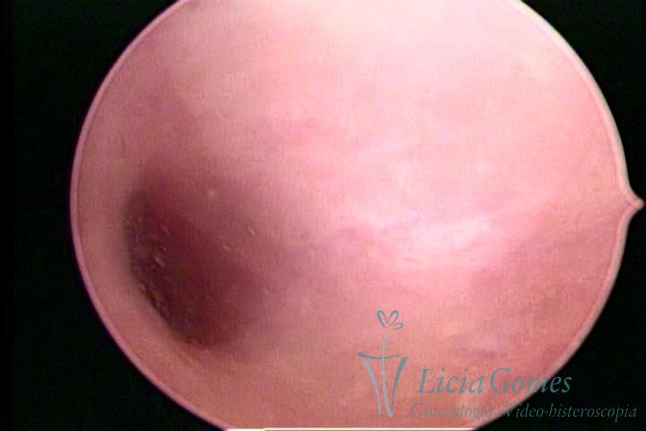 54
54 55
55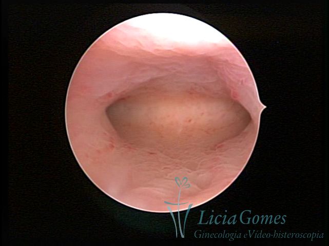 56
56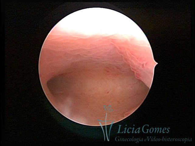 57
57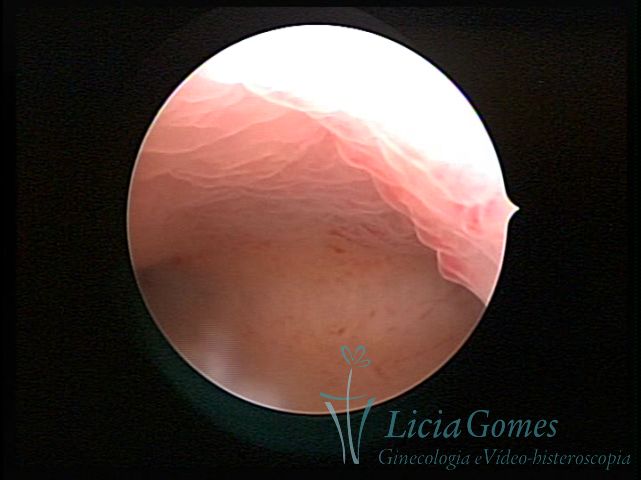 58
58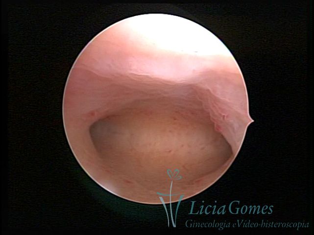 59
59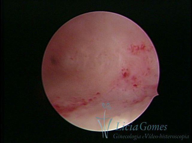 60
60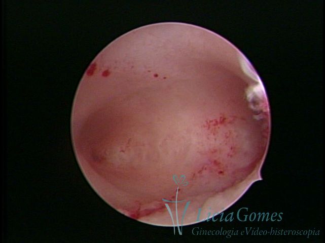 61
61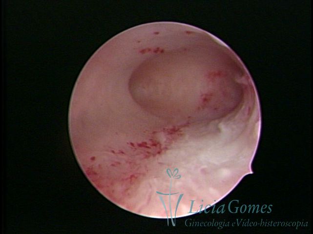 62
62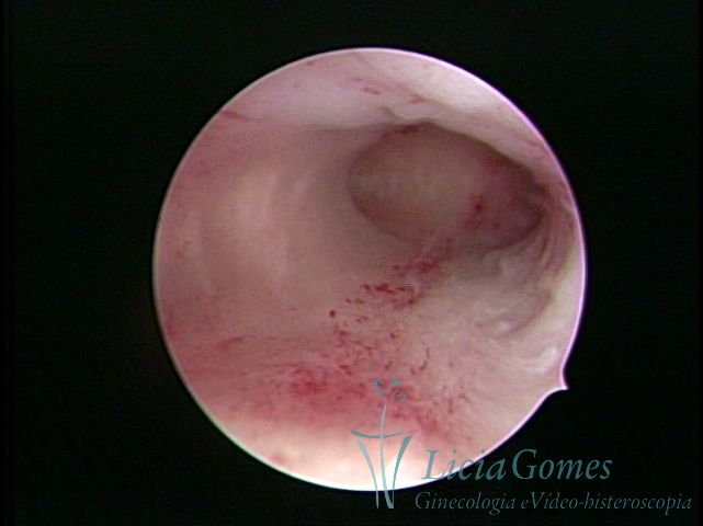 63
63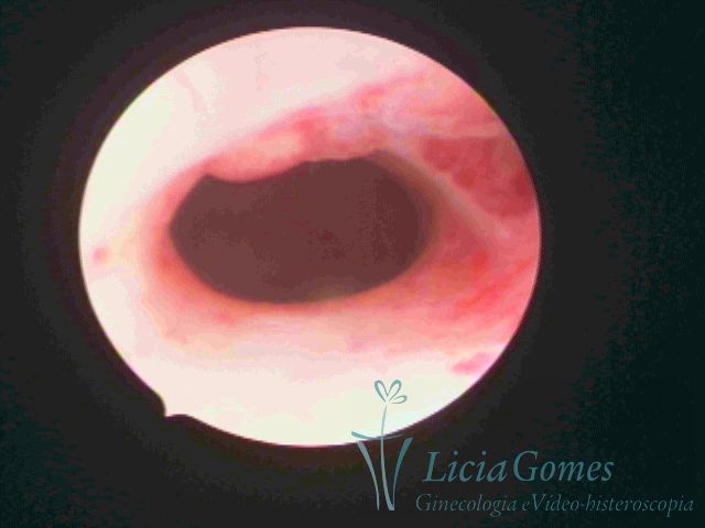 64
64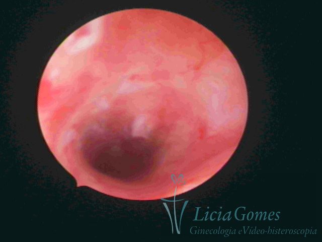 65
65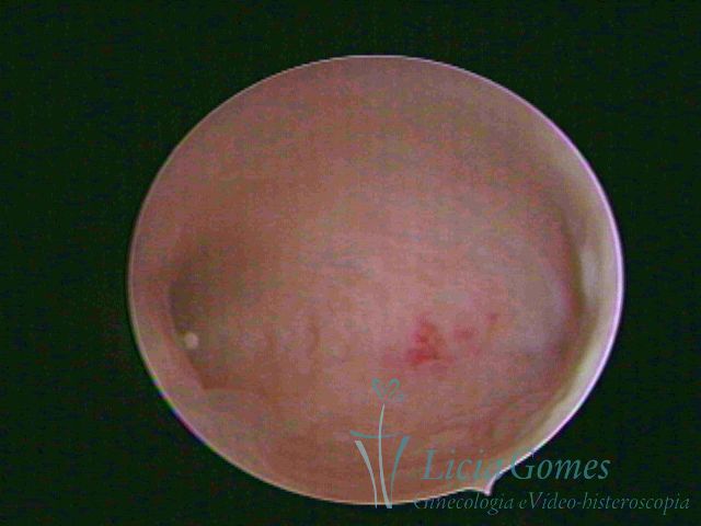 66
66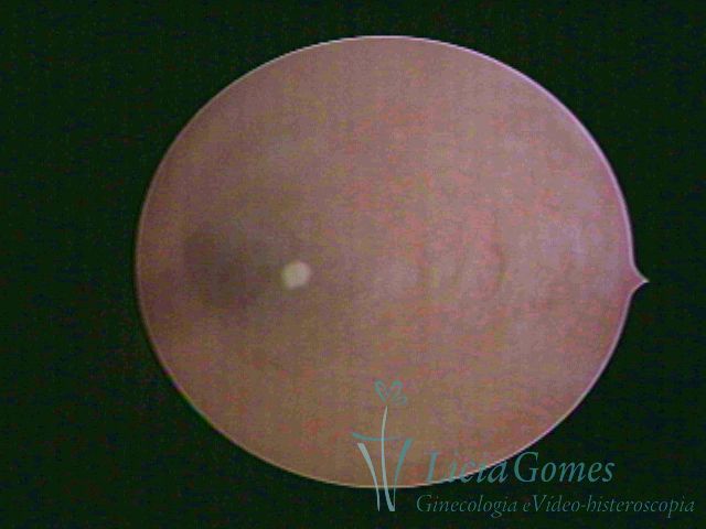 67
67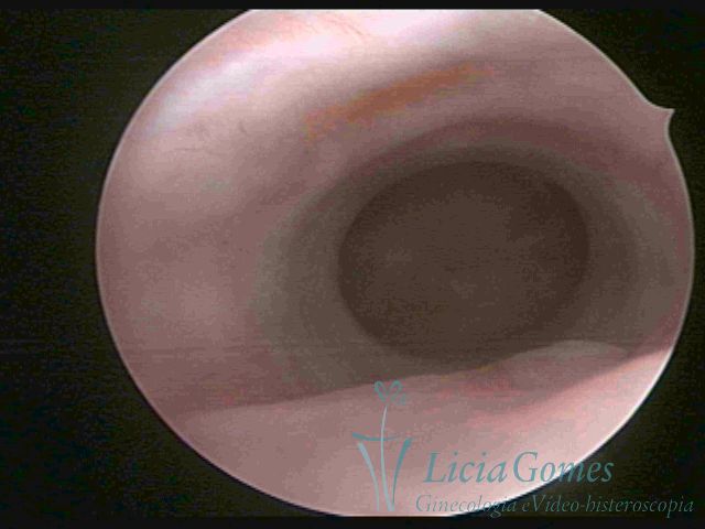 68
68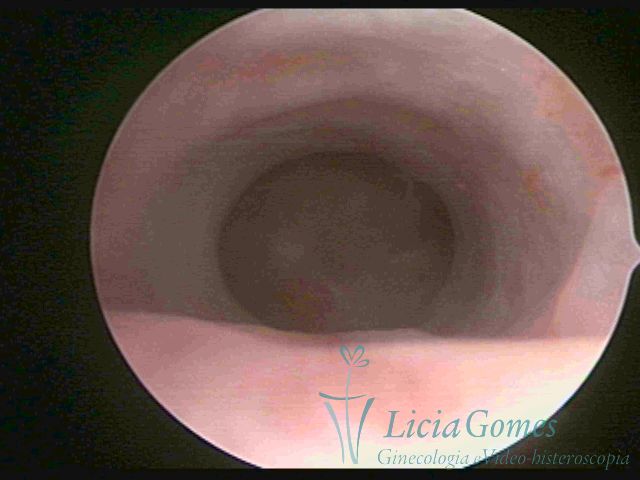 69
69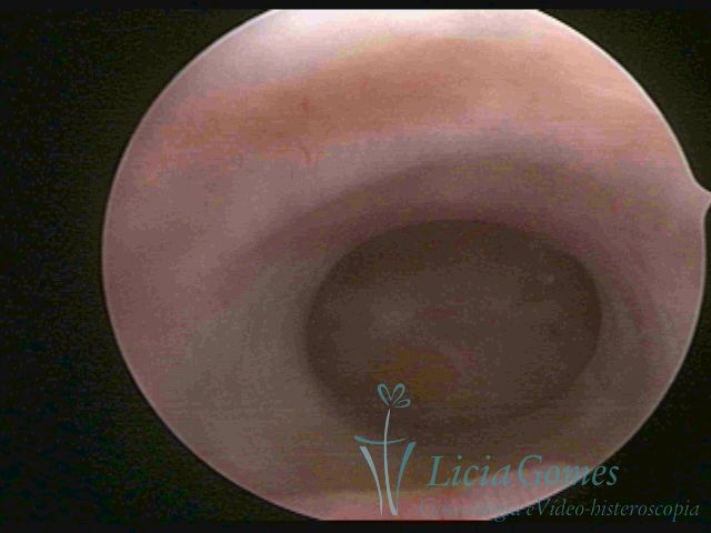 70
70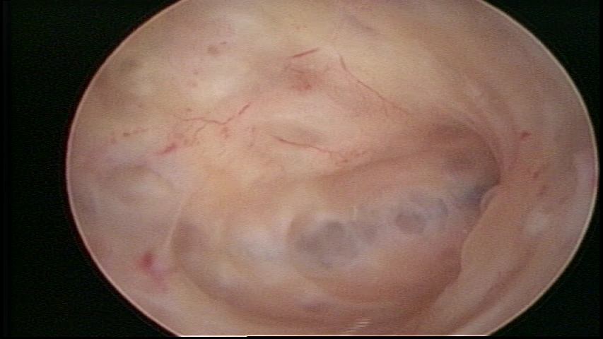 71
71