Brasil
 Global
NORMAL CERVICAL CANAL
The cervical canal is different during menacme and menopause. In hysteroscopy, the cervical canal is commonly divided into three parts:
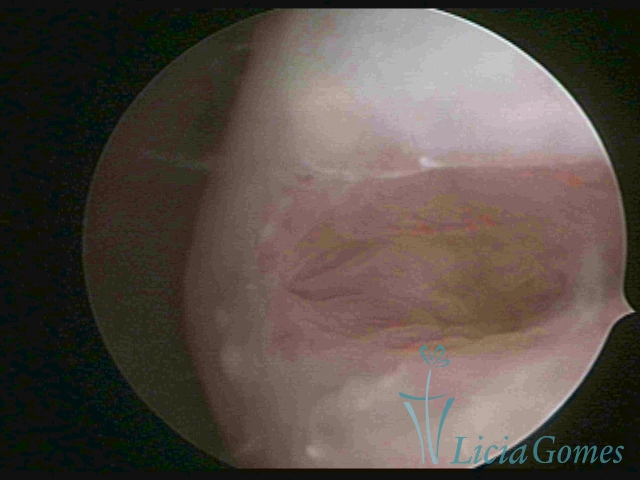
First part or proximal section or lower section:
During the proliferative phase, a light, crystalline mucus with a low adherence to the scope is found. The crypts and buds are a little swelled and vascularized, microvesicular, resembling grape clusters.
Second part or middle section
In the middle section of the cervical canal, the details of the buds are lost. It is possible to view creases and crypts. Longitudinal grooves are frequently observed. They are the most compact tissues presenting the most vascularized surface, whose vessels follow its passageway.
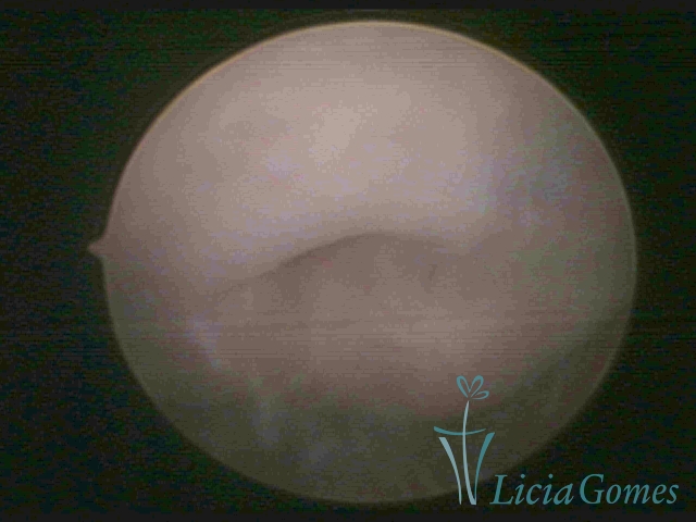
Third part or upper section
Presents the mucosa with a smooth, poorly vascularized surface, to the internal orifice
 1
1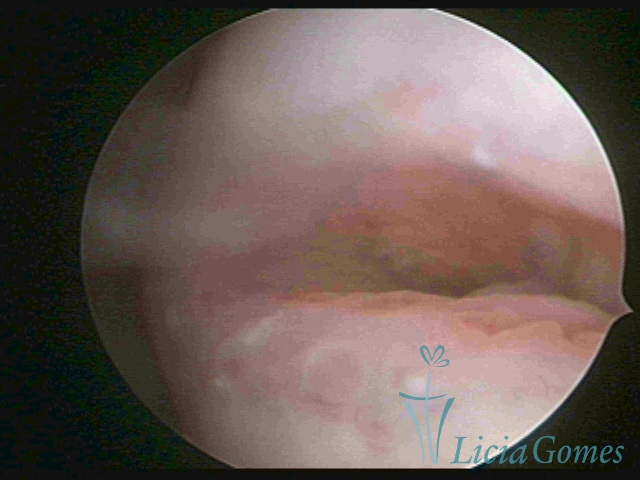 2
2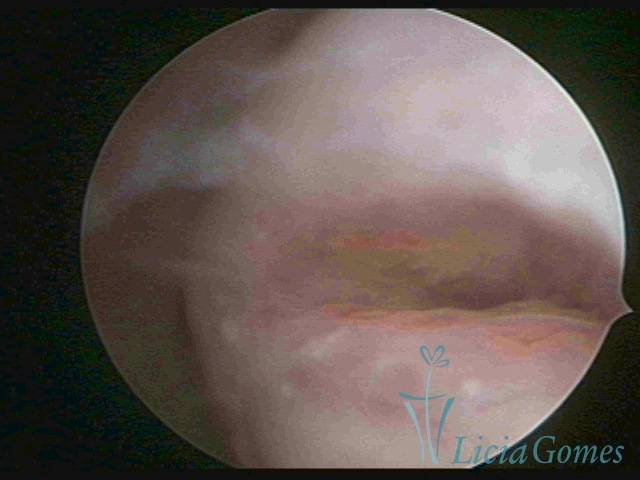 3
3 4
4 5
5 6
6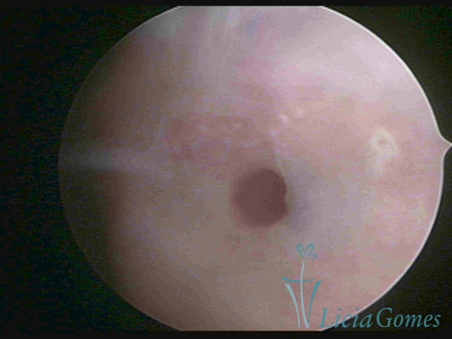 7
7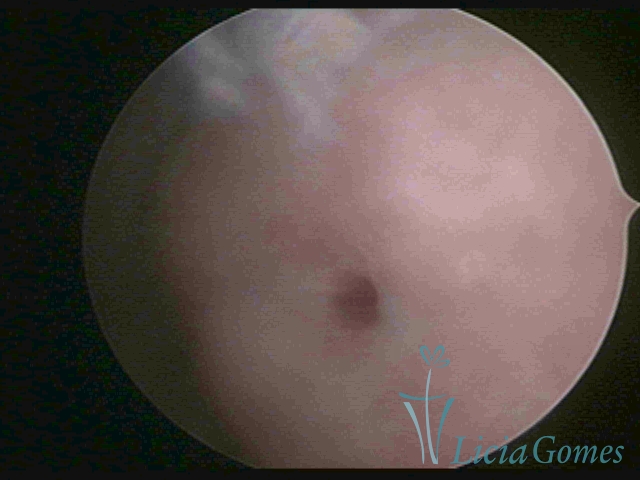 8
8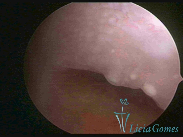 9
9 10
10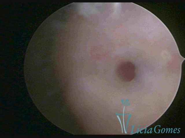 11
11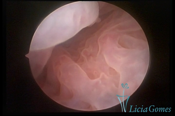 12
12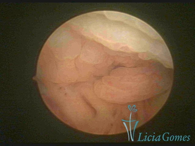 13
13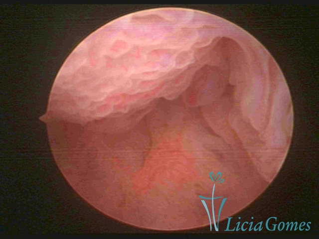 14
14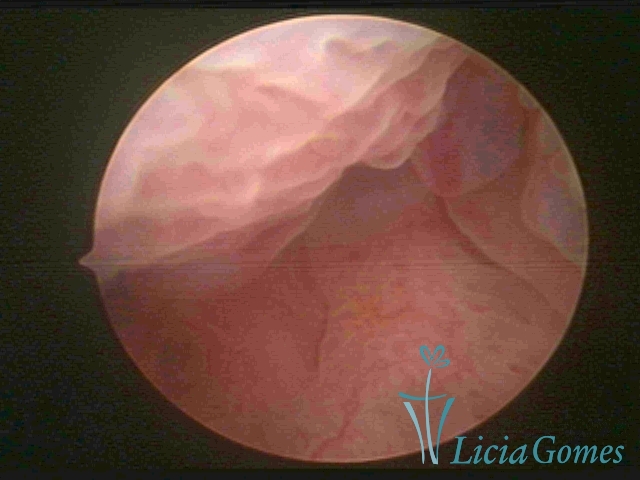 15
15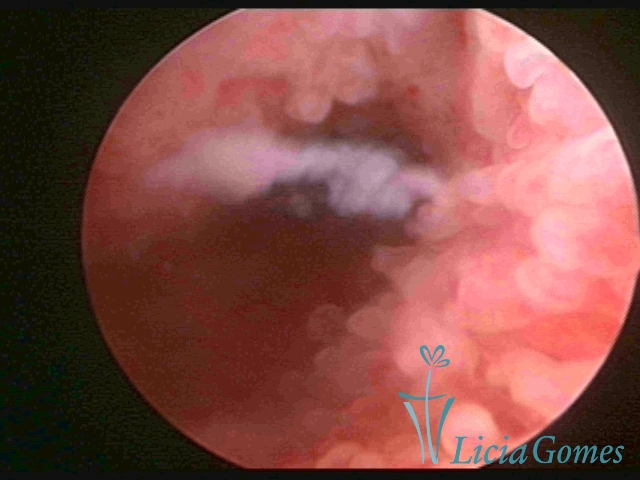 16
16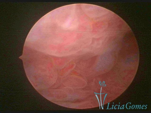 17
17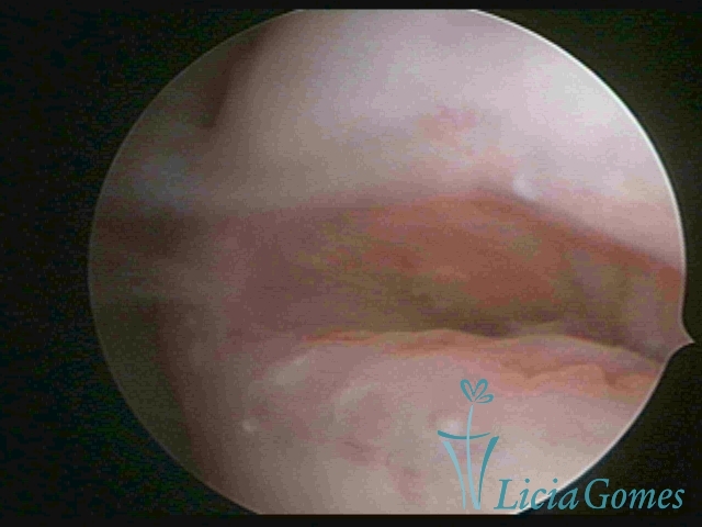 18
18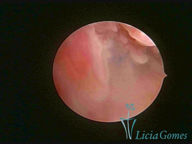 19
19 20
20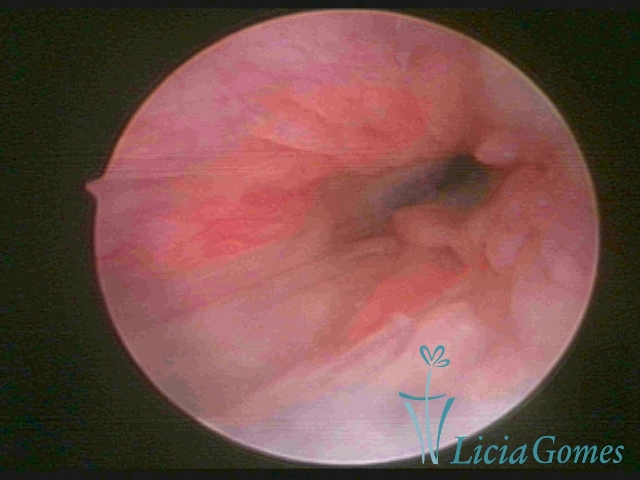 21
21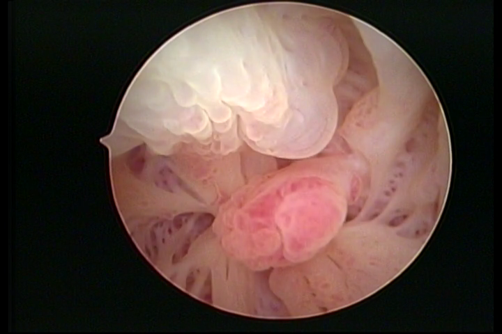 22
22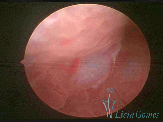 23
23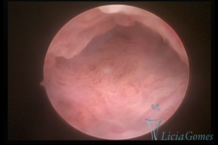 24
24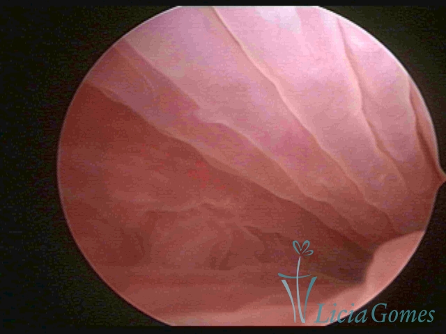 25
25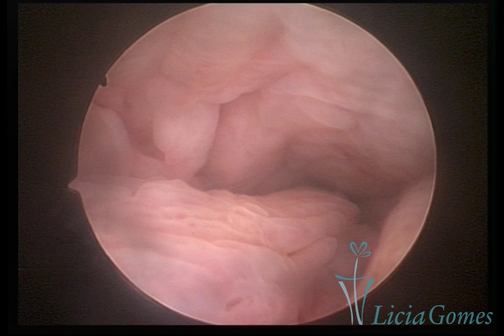 26
26 27
27 28
28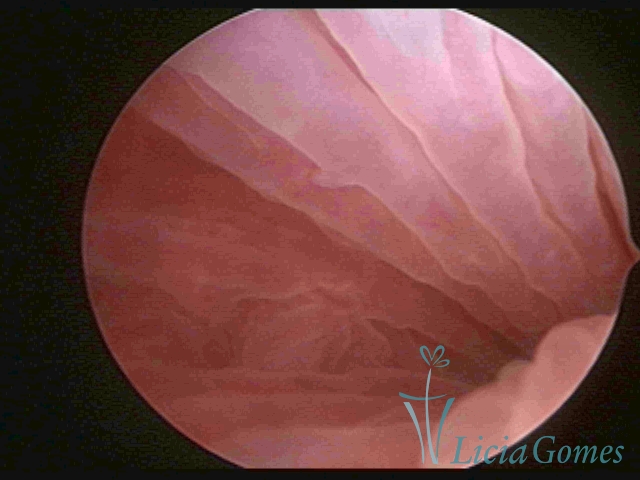 29
29 30
30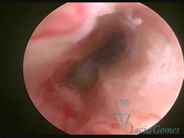 31
31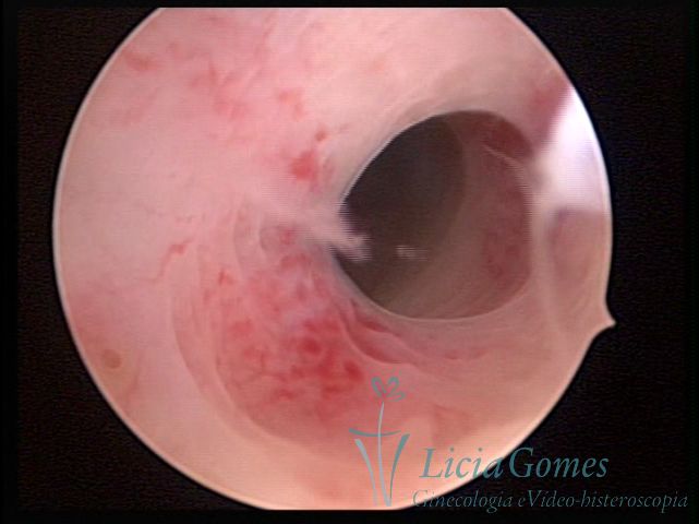 32
32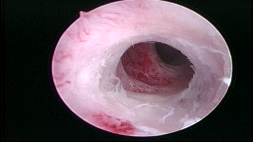 33
33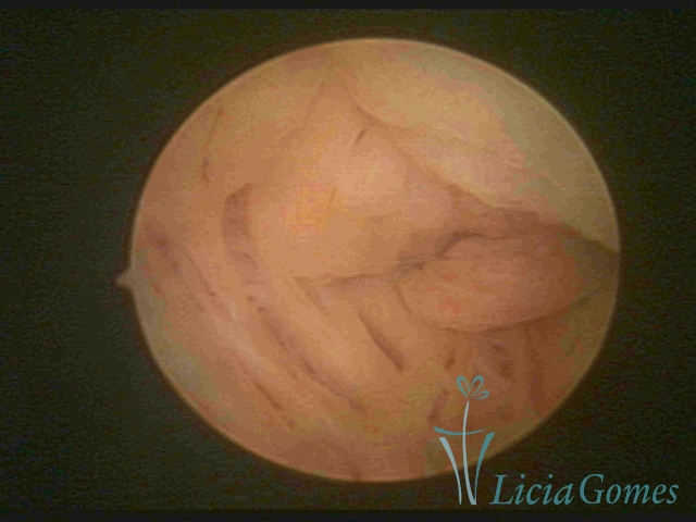 34
34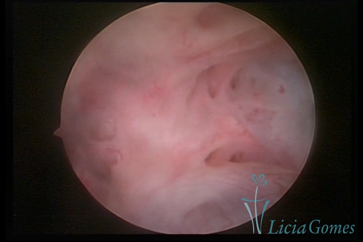 35
35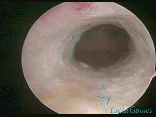 36
36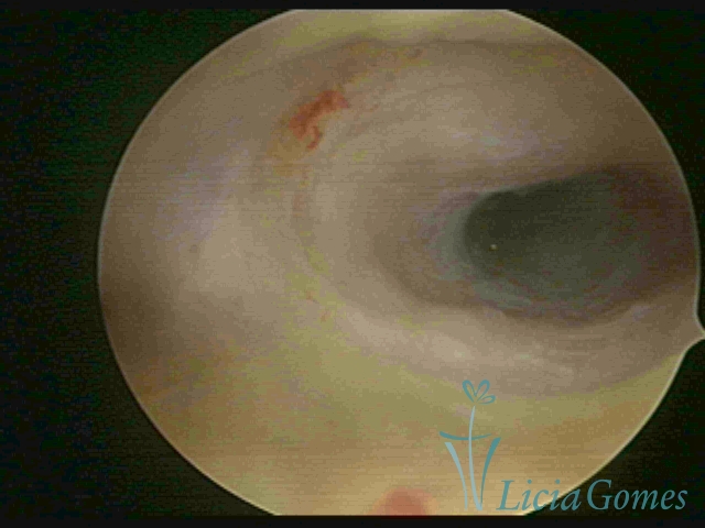 37
37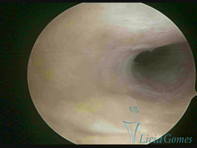 38
38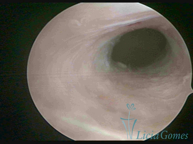 39
39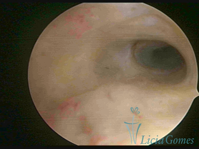 40
40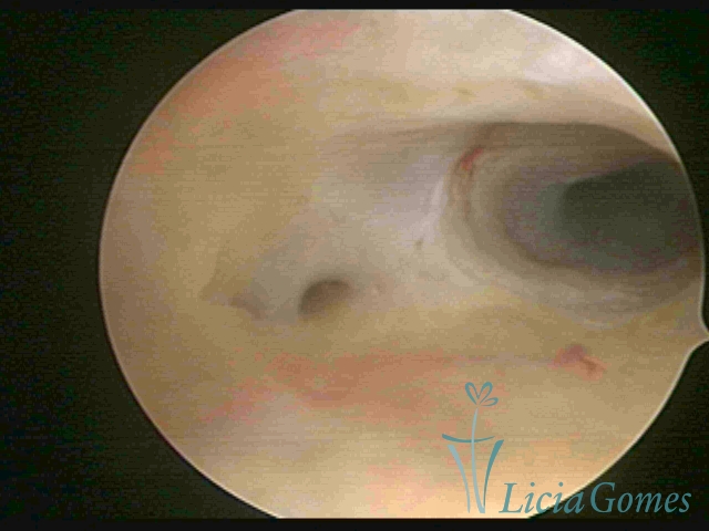 41
41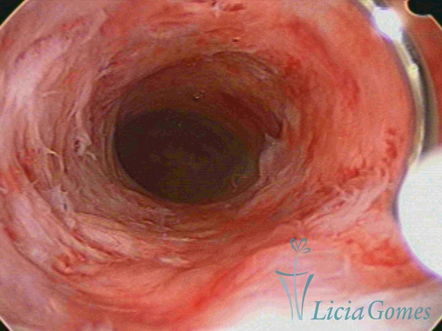 42
42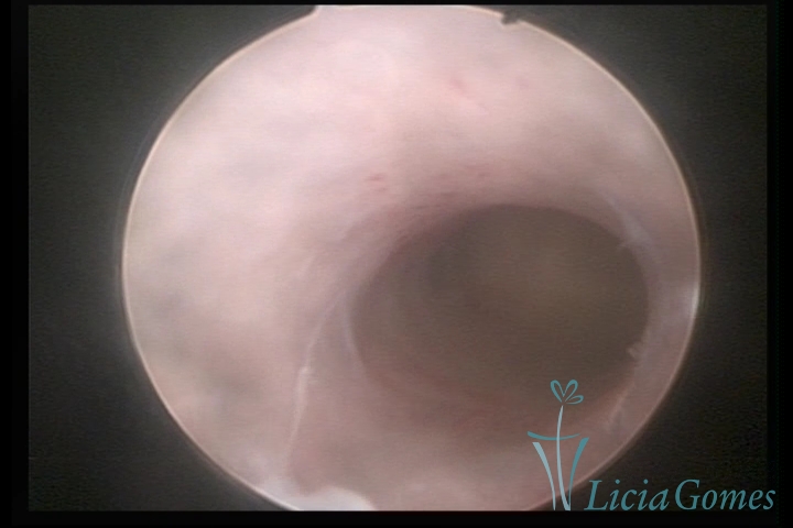 43
43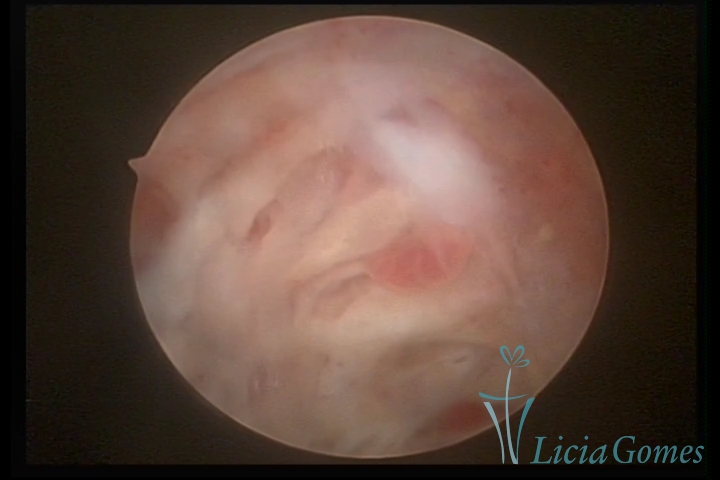 44
44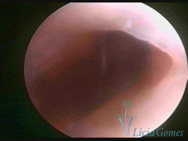 45
45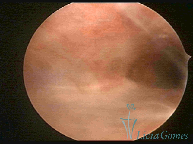 46
46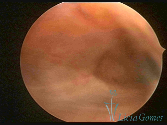 47
47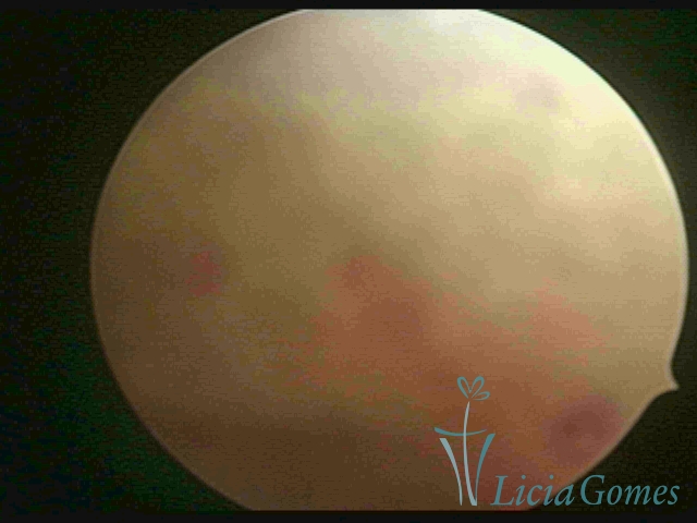 48
48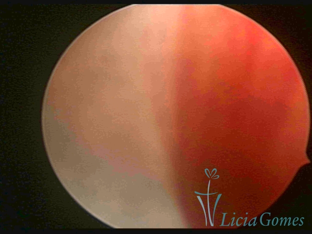 47
47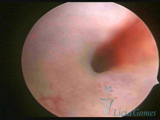 49
49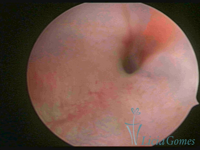 50
50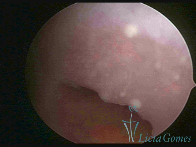 51
51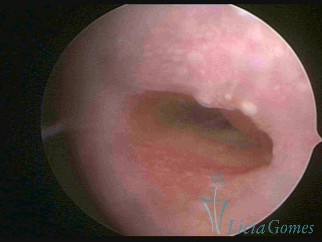 52
52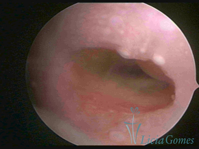 53
53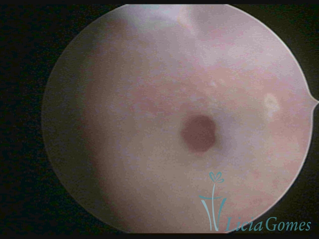 54
54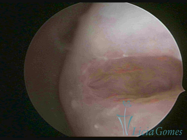 55
55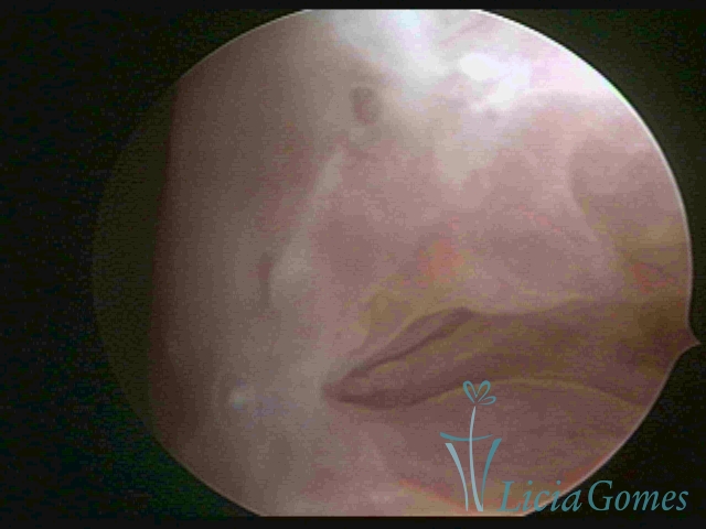 56
56 57
57