Brasil
 Global
Malignant neoplasia
NEOPLASIAS OF THE UTERINE BODY
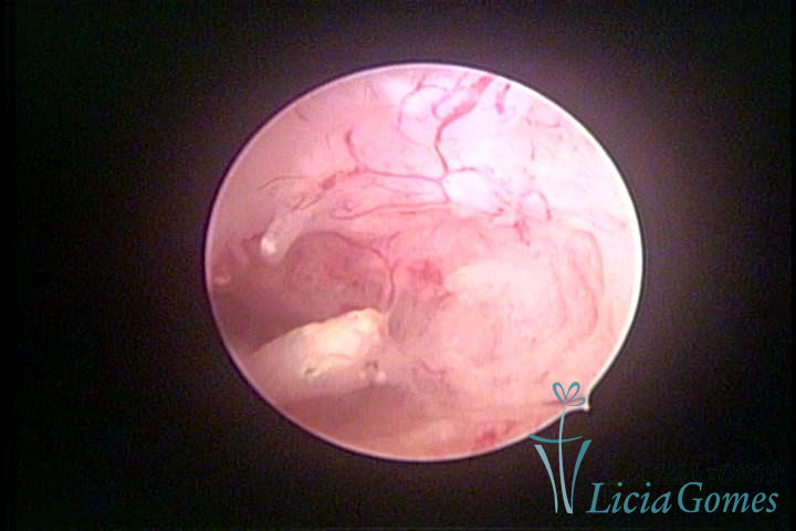
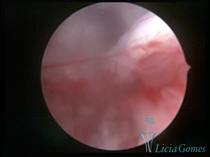
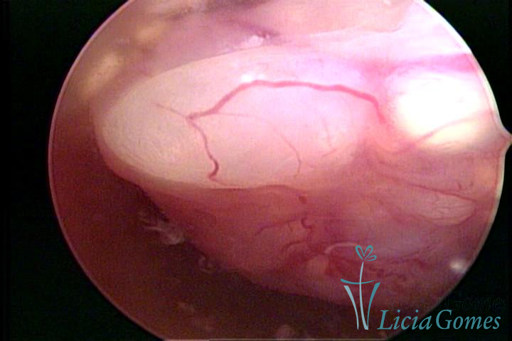
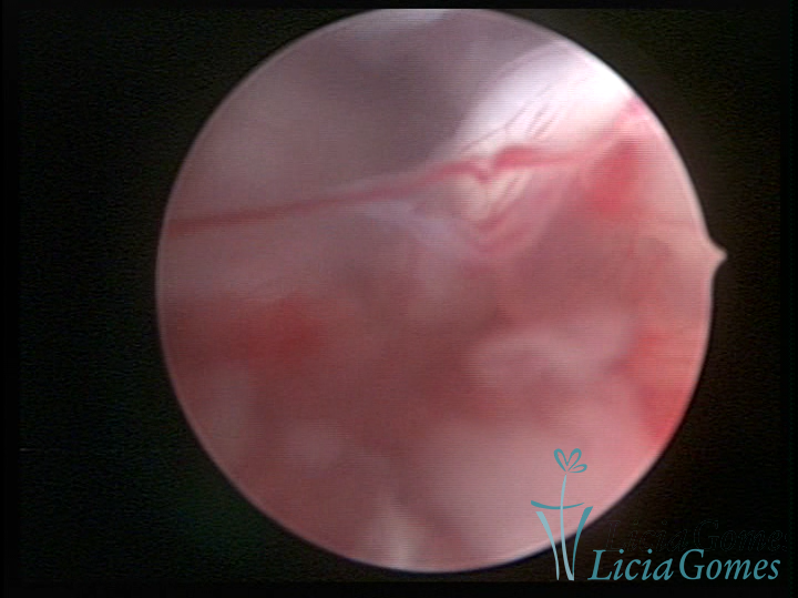
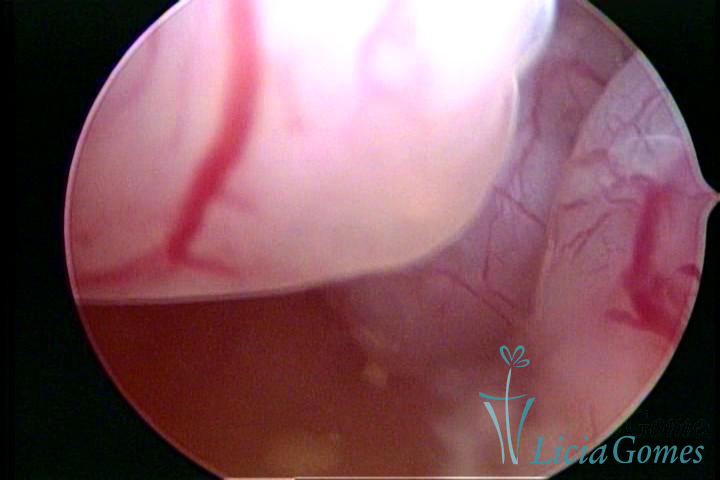
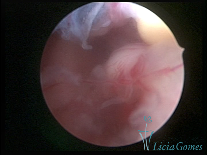
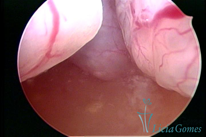
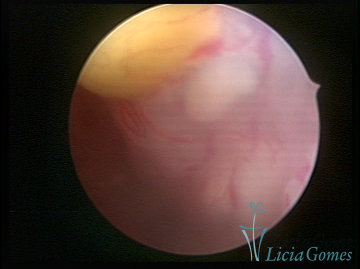
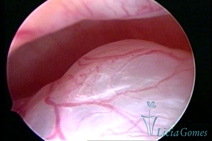
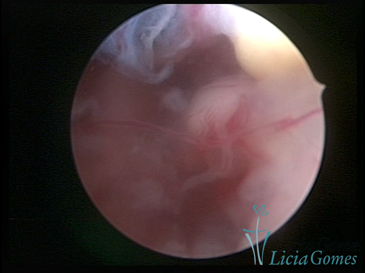
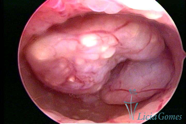
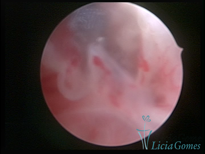
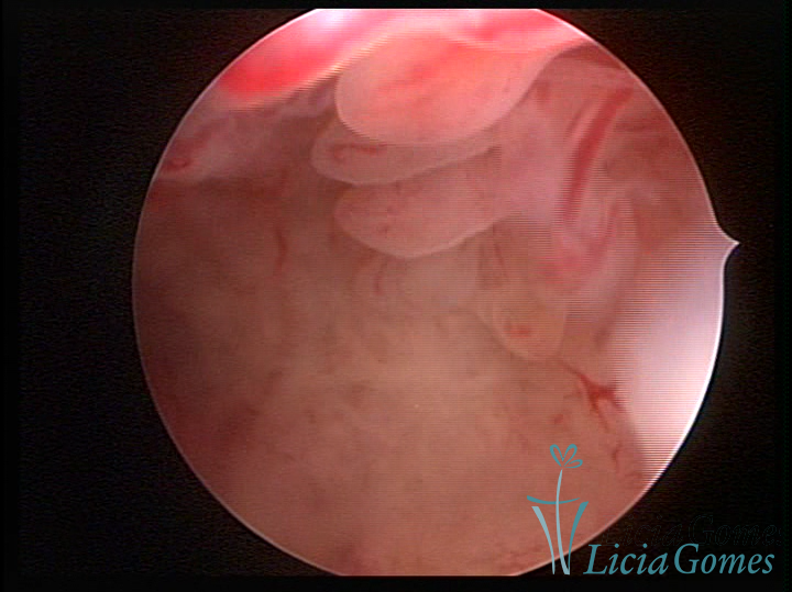
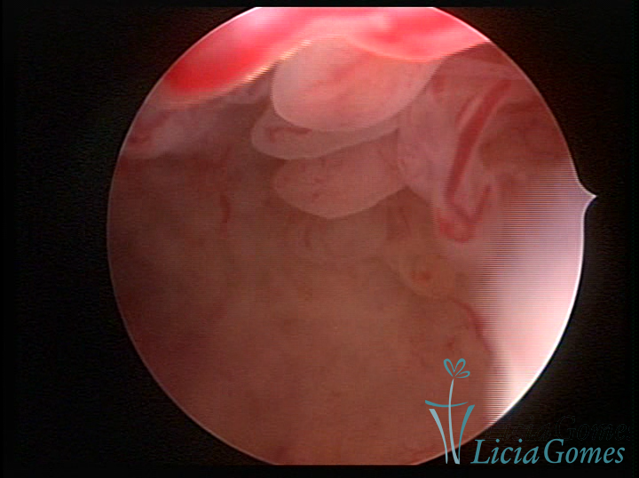
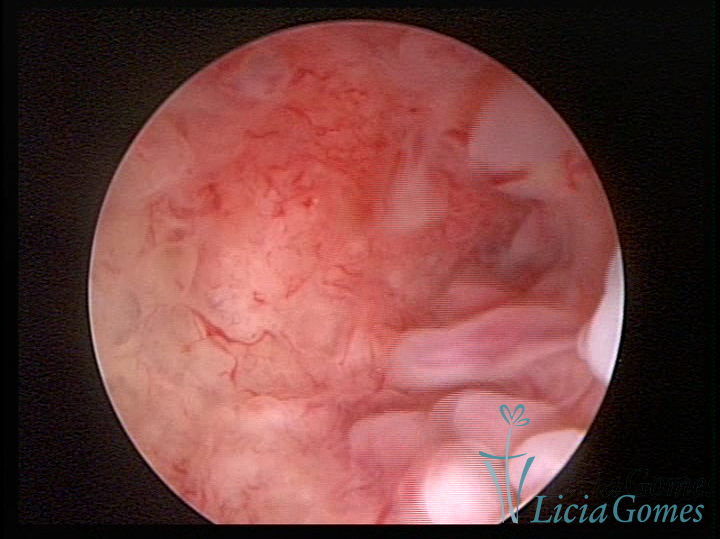
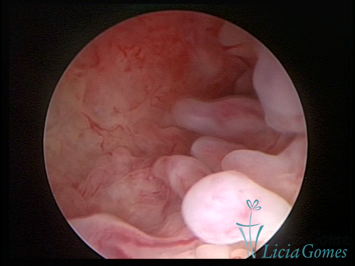
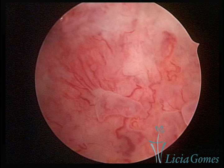
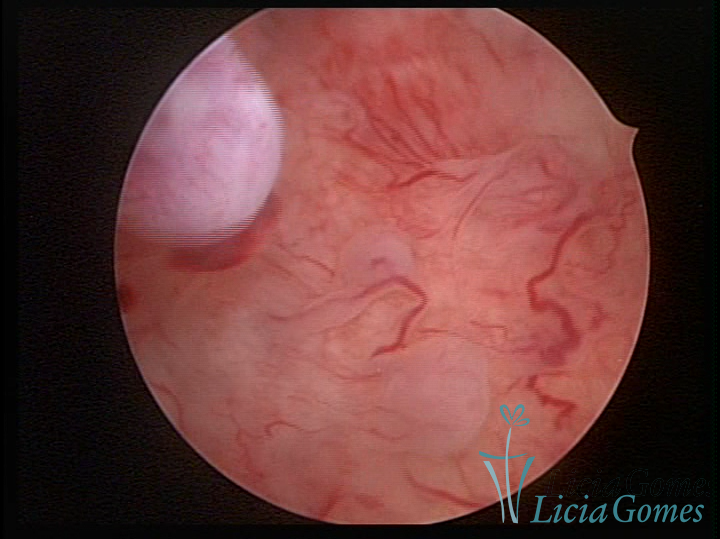
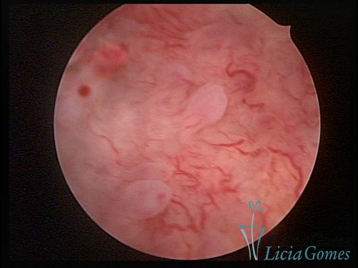
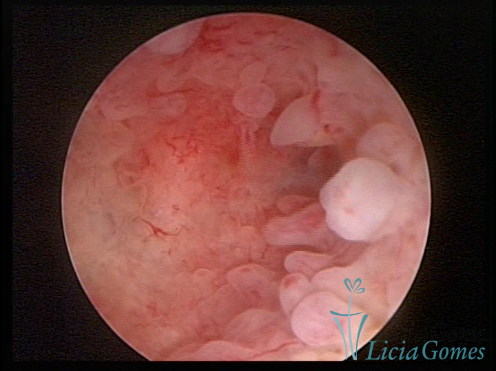
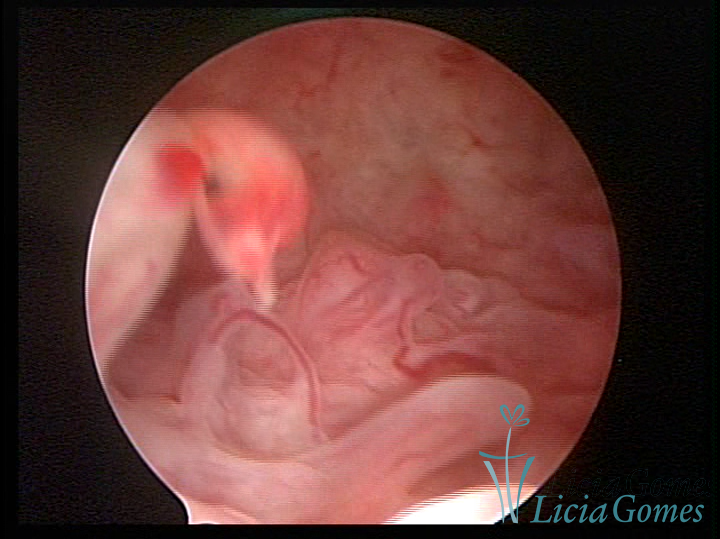
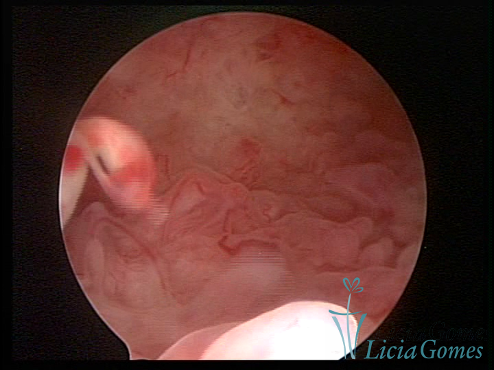
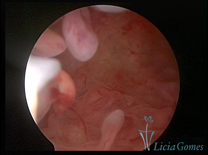
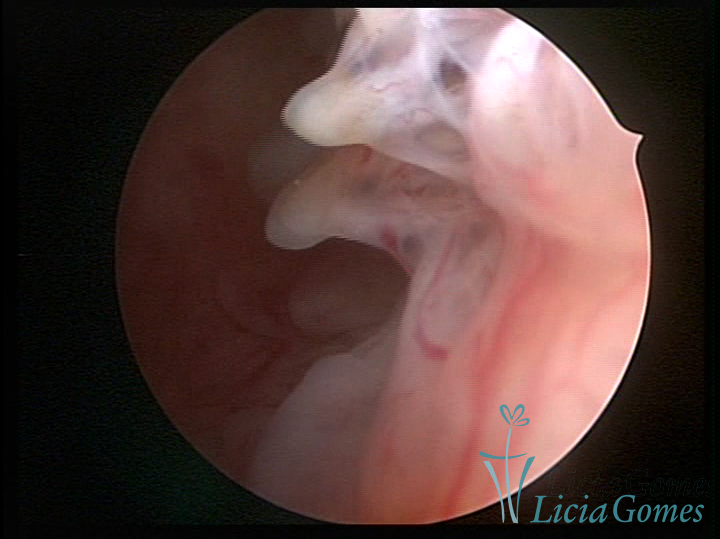
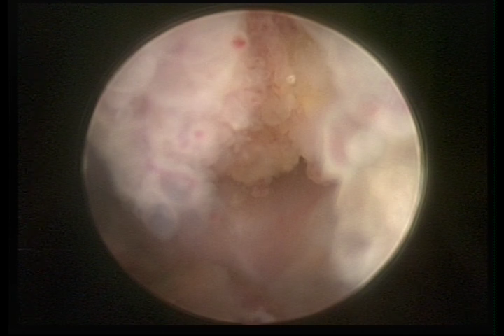
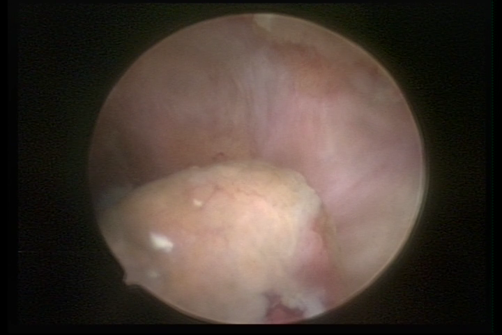
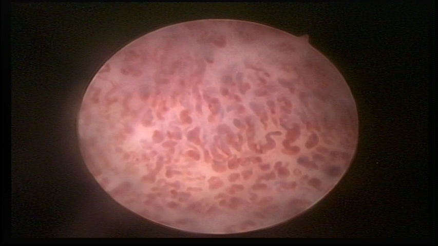
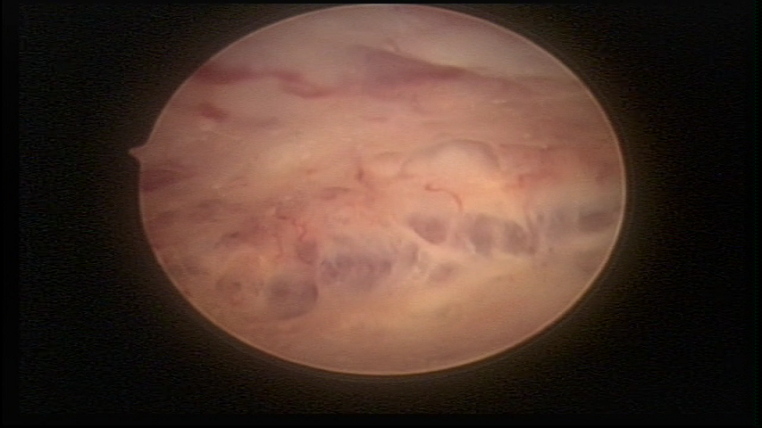
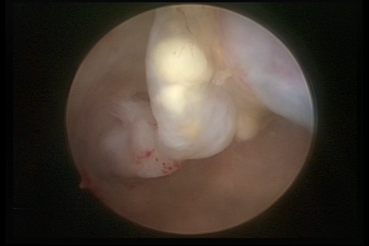
Hysteroscopy can analyze the characteristics on the endometrial surface, not being able to evaluate the depth or the degree of myometrial invasion.
However, it may be able to detect focal, regional or diffuse lesions.
It is important to highlight that microscopy (Histopathological) is responsible for the differential diagnosis of Hyperplasia with atypia and Adenocarcinoma of the endometrium or another type of endometrial neoplasia.
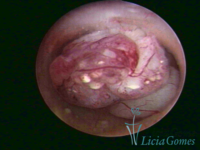
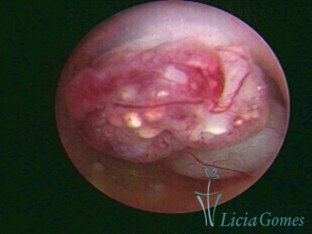
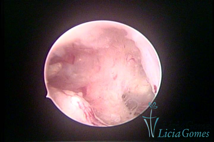
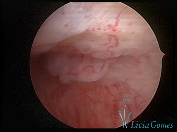
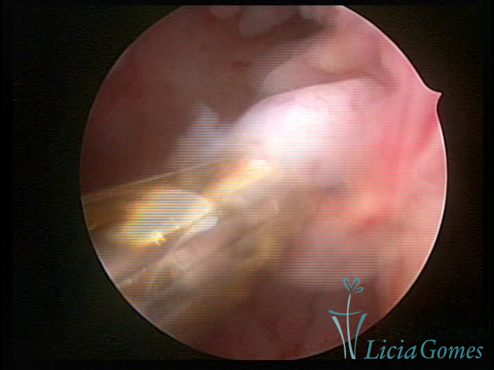
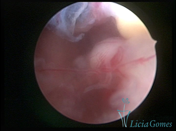
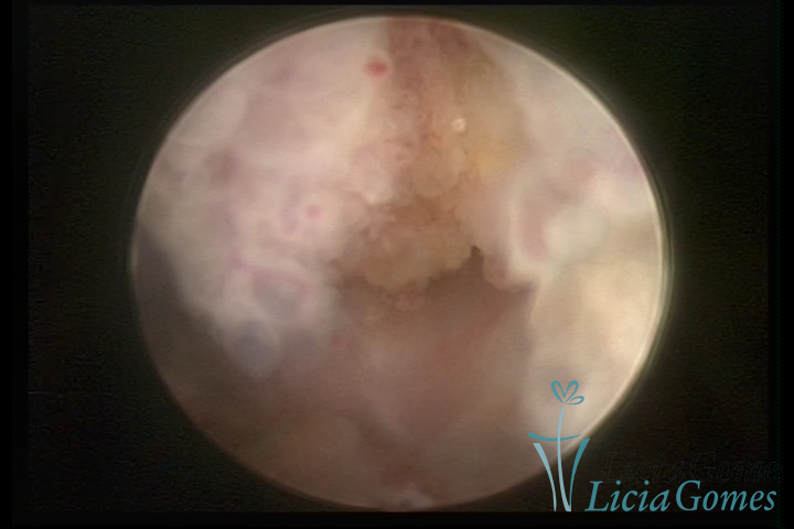
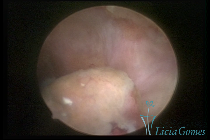
It may present a variable range of macroscopic aspects, with a pseudolopypoid aspect; resembling cerebroid tissue or presenting a decidual reaction; superficial vascularization is more evident presenting vessels in the shape of corkscrew or spirals also viewing the vascularization with atypias, with an increase on the thickness of the superficial vessels, tissue in necrosis and small dendrites (papillomatous) may be found.